
This is an old revision of the document!
Table of Contents

|
Location: Room P2-A-22 ( |
 Zeiss LSM 880 Booking
 Zeiss LSM 880 Usage Statistics
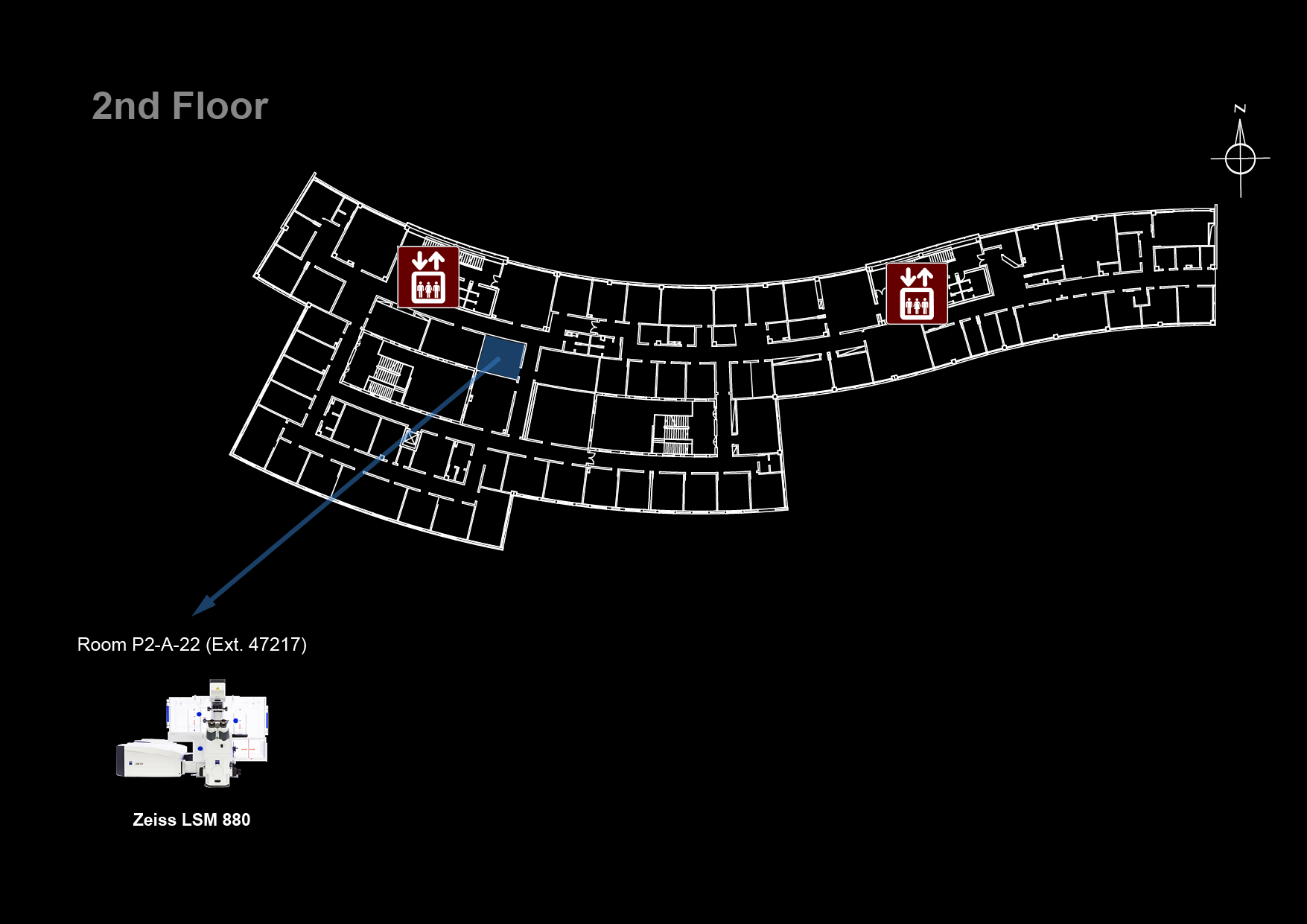
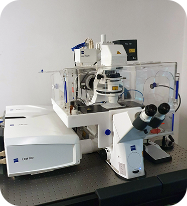
Microscope overview
 The Zeiss LSM 880 with Airyscan is a confocal point-scanning microscope able to generate high-resolution three-dimensional images of thick specimens with high sensitivity, high speed and low photodamage. It is an inverted microscope especially suitable for live cell imaging and photobleaching experiments, equipped with a large size incubator for temperature control and CO2 supply and with Definite Focus for hardware focus control during long time-lapse acquisitions. The stage is motorized and furthermore equipped with a piezo for Z displacement so fast 4D imaging is possible in multiple stage positions. Its Airyscan 32 channel area detector is able to collect all light from an Airy pattern simultaneously, allowing for increased resolution (140 nm laterally and 400 nm axially, at 488 nm). It can also be used in Fast Mode, which enables parallel excitation and detection of four image pixels. The result is a speed improvement by a factor of four, while maintaining the sensitivity of Airyscan and 1.5x resolution improvement. Its scanning unit further includes two PMT detectors (one of them cooled) as well as a GaAsP detector for increased sensitivity (45% QE compared to ~25% QE for conventional PMT). It is equipped with lasers from violet to far red (405, 458, 488, 514, 561, 594 and 633 nm excitation wavelengths).
 With this system you can perform optical sectioning of fluorescent samples which are too thick for a widefield system such as the Zeiss Cell Observer or the Zeiss Axiovert 200M. Image resolution with Airyscan is higher than the Zeiss LSM 710 and detection sensitivity is also higher than the spinning disks 3i Marianas SDC and Zeiss Cell Observer SD. If your personal computer cannot handle all the data you collected, check out the Big Guy or Colossus.
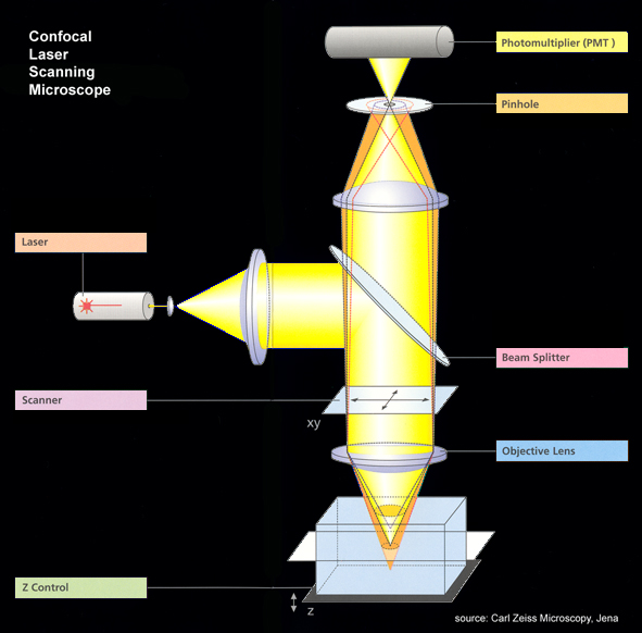
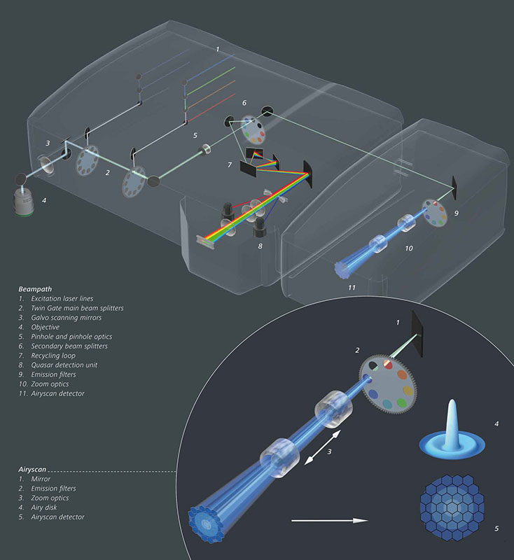
![]() Click here to see the system beam path in higher detail
Click here to see the system beam path in higher detail
![]() Data files older than 1 month will be automatically deleted on this system, please copy your data to the iMM server using the desktop link.
Data files older than 1 month will be automatically deleted on this system, please copy your data to the iMM server using the desktop link.
Additional information
Booking Rules
- Users can book at maximum 8 hours per week
- This usage restriction does not apply for weekends and for working days before 9:00 and after 21:00
- Exceptions to these rules require approval from José Rino.
System components
LASERs
| Laser Unit | Wavelength | Maximum Power | Current Status |
|---|---|---|---|
| Diode 405-30 | 405 nm | 30 mW | ok |
| Argon | 458, 488 and 514 nm | 25 mW | ok |
| DPSS 561-20 | 561 nm | 20 mW | ok |
| HeNe594 | 594 nm | 2 mW | ok |
| HeNe633 | 633 nm | 5 mW | ok |
Objectives
| Magnification | Model | Immersion | NA | WD (mm) | Reference |
|---|---|---|---|---|---|
| 10x | EC Plan-Neofluar | Air | 0.30 | 5.20 | 420340-9901-000 |
| 20x | Plan-Apochromat | Air | 0.80 | 0.55 | 420650-9901-000 |
| 40x | C-Apochromat Corr | Water | 1.20 | 0.28 | 421767-9970-000 |
| 63x | Plan-Apochromat DIC | Oil | 1.40 | 0.19 | 420782-9900-799 |
Upon request:
| Magnification | Model | Immersion | NA | WD (mm) | Reference |
|---|---|---|---|---|---|
| 25x | LCI Plan-Neofluar Corr DIC | Oil/Glyc/W | 0.80 | 0.21 | 420852-9972-000 |
| 40x | Plan-Apochromat Corr | Dry | 0.95 | 0.25 | 420660-9970-000 |
| 100x | α Plan-Apochromat DIC | Oil | 1.46 | 0.11 | 420792-9800-000 |
Filtersets (Ocular)
Microscope Turn On Procedure
- Turn on the
MAIN SWITCH - Turn on the
SYSTEMS/PCswitch

- Turn on the computer
- Login in to Windows (Bioimaging User)
- Turn on the
COMPONENTSswitch - Check that the computer is connected to an Unidentified network (click on the network icon in the taskbar)
- Start the ZEN Black software
Warning: If you need to change the stage adapter, please contact the Bioimaging Unit (imm-bioimaging@medicina.ulisboa.pt | ![]() 47222)
47222)
Microscope Turn Off Procedure
If there is another user for this microscope in the next hour:
- Exit the ZEN software, leave the lasers on
- Log off the computer
- Clean up immersion objectives
Else:
- Turn off the lasers on ZEN
- Clean up immersion objectives
- Exit the ZEN software
- Shutdown the computer
- Turn off the two switches
SYSTEMS/PCandCOMPONENTS - Wait 5 min. for Ar laser to cool down
- Turn off the
MAIN SWITCH