
Table of Contents

|
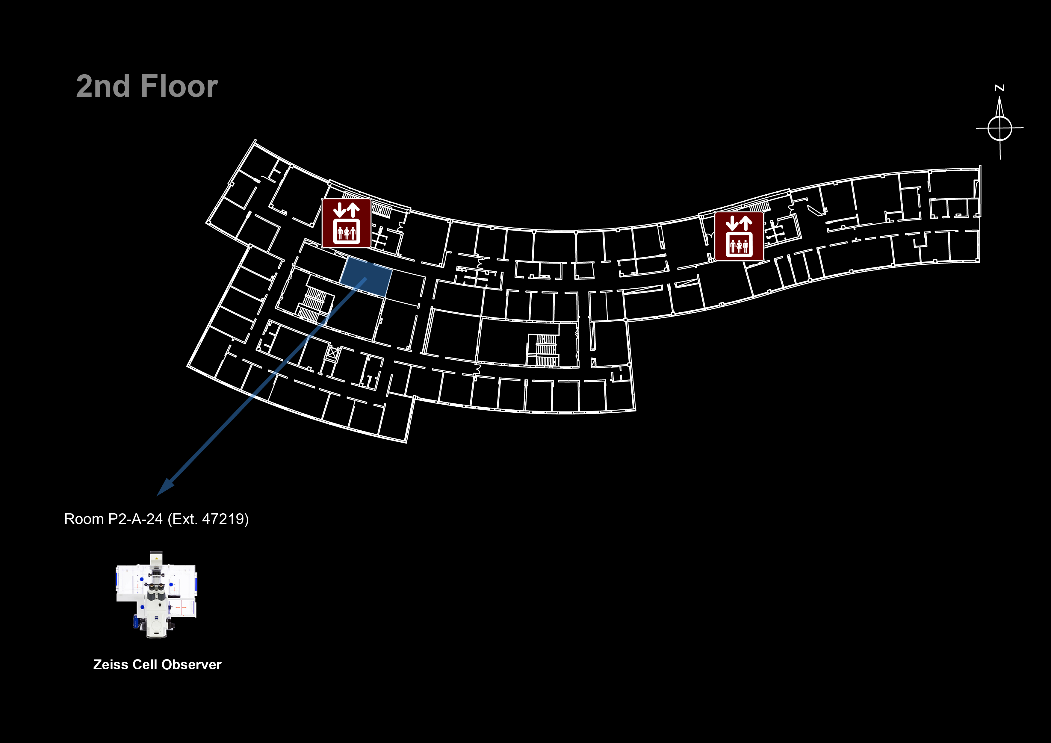
Location: [Lisbon] Room P2-A-24 ( |
 47219)
Microscope overview
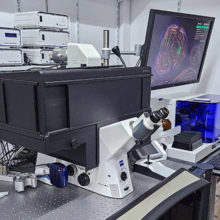
 The Zeiss Cell Observer is a fully motorized inverted widefield fluorescence microscope ideal for live-cell imaging with fast acquisition speeds. It is equipped with a large cage incubator and a stage top incubator for temperature control and CO2 supply and with Definite Focus for hardware focus control during long time-lapse acquisitions. Using the ZEN software you can control the motorized filters, shutters and stage to simultaneously set up multi-color time-lapse imaging experiments in multiple stage positions. The stage is furthermore equipped with a piezo for fast Z displacement. You can choose between a HXP metal halide lamp or fast-switching LEDs for illumination. Its cooled Axiocam 506m camera allows you to capture weak fluorescent signals and minimize photobleaching/photodamage in light sensitive samples. It is also equipped with a high sensitivity Photometrics Prime BSI Express sCMOS camera for high resolution imaging with maximized signal detection.
- Microscope: Zeiss Axio Observer
- LED Illumination: Zeiss Colibri 7
- Camera (left port): Axiocam 506 mono CCD
- Camera (right port): Photometrics Prime BSI Express sCMOS
 This system is also set up to work together with the AKOYA PhenoCycler fluidics instrument for automated iterative imaging cycles of whole slide tissues for Spatial Biology applications, such as image-based quantification of biomarkers to predict immunotherapy response, or quantification of cells by lineage and variant with single-cell resolution in the context of an intact tissue sample.
 Data files older than 15 days will be automatically deleted on this system, please copy your data to the GIMM server using the desktop link.
System components
LEDs
| Line | Wavelength |
|---|---|
| UV | 385 nm |
| B | 475 nm |
| G | 555 nm |
| Y | 590 nm |
| R | 630 nm |
Filtersets
| Position | Filterset | Excitation | Dichroic | Emission | Reference |
|---|---|---|---|---|---|
| 1 | Green | 450-490 nm | 495 nm | 500-550 nm | FS38HE |
| 2 | Orange | 538-562 nm | 570 nm | 570-640 nm | FS43HE |
| 3 | Red | 625-655 nm | 660 nm | 665-715 nm | FS50 |
| 4 | Analyzer DIC | ||||
| 5 | QUAD | 370-400 nm | 405 nm | 410-440 nm | FS90HE |
| 450-488 nm | 493 nm | 499-529 nm | |||
| 540-570 nm | 575 nm | 580-605 nm | |||
| 615-648 nm | 653 nm | 659-759 nm | |||
| 6 | DAPI | 335-383 nm | 395 nm | 420-470 nm | FS49 |
Objectives
| Magnification | Model | Immersion | NA | WD (mm) | Reference |
|---|---|---|---|---|---|
| 10x | EC Plan-Neofluar Ph1 | Air | 0.30 | 5.20 | 420341-9911-000 |
| 20x | Plan-Apochromat | Air | 0.80 | 0.55 | 420650-9901-000 |
| 20x | EC Plan-Neofluar Ph 2 | Air | 0.50 | 2.00 | 440341-9904-000 |
| 40x | EC Plan-NeoFluar | Air | 0.75 | 0.71 | 420360-9900-000 |
| 63x | Plan-Apochromat DIC | Oil | 1.40 | 0.19 | 420782-9900-000 |
| 63x | Plan-Apochromat Ph3 | Oil | 1.40 | 0.19 | 420781-9910-000 |
Upon request:
| Magnification | Model | Immersion | NA | WD (mm) | Reference |
|---|---|---|---|---|---|
| 1.25x | EC Epiplan-Neofluar | Air | 0.035 | 4.00 | 422310-9900-000 |
| 5x | EC Plan-NeoFluar | Air | 0.16 | 18.50 | 420330-9901-000 |
| 10x | EC Plan-Neofluar | Air | 0.30 | 5.20 | 420340-9901-000 |
| 25x | LCI Plan-Neofluar Corr DIC | Oil/Glyc/W | 0.80 | 0.21 | 420852-9972-000 |
| 40x | Plan-Apochromat Corr | Air | 0.95 | 0.25 | 420660-9970-000 |
| 40x | Plan-Neofluar Ph3 | Oil | 1.30 | 0.20 | 440451-9903-000 |
| 100x | Plan-Apochromat DIC | Oil | 1.40 | 0.17 | 440782-9902-000 |
Camera
| Model | Type | Frame Size | Pixel Size (µm) |
|---|---|---|---|
| Axiocam 506 mono | CCD | 2752 x 2208 | 4.54 x 4.54 |
System Turn On Procedures
- Turn on the HXP 120 fluorescent light source (if used)

- Turn on the temperature and CO2 controller - white power strip (if you need them).
- If you need CO2, open the Cell Observer CO2 valve
- Turn on the motorized stage controller (1 on remote control)

- Turn on the microscope power supply (2 on remote control)

- Turn on the computer (if needed)
- Login in to Windows with your Agendo credentials
- Start the ZEN software
- Select HXP or LED Workspace in ZEN (top left)

System Turn Off procedures
If there is another user for this microscope is the next hour:
- Exit the ZEN software
- Log off the computer
- Leave the fluorescent light source on
- Clean up immersion objectives
Else:
- Exit the ZEN software
- Turn off the computer
- Clean up immersion objectives
- Turn off the microscope power supply (2 on remote control)
- Turn off the motorized stage controller (1 on remote control)
- Turn off the Temperature and CO2 controller - white power strip (if used)
- Close the Cell Observer CO2 valve (if used)
- Turn off the HXP 120 fluorescent light source (if used)