User Tools
This is an old revision of the document!
Table of Contents

|
Location: Room P2-A-24 ( |
 Zeiss Axiovert 200M Booking
 Zeiss Axiovert 200M Usage Statistics
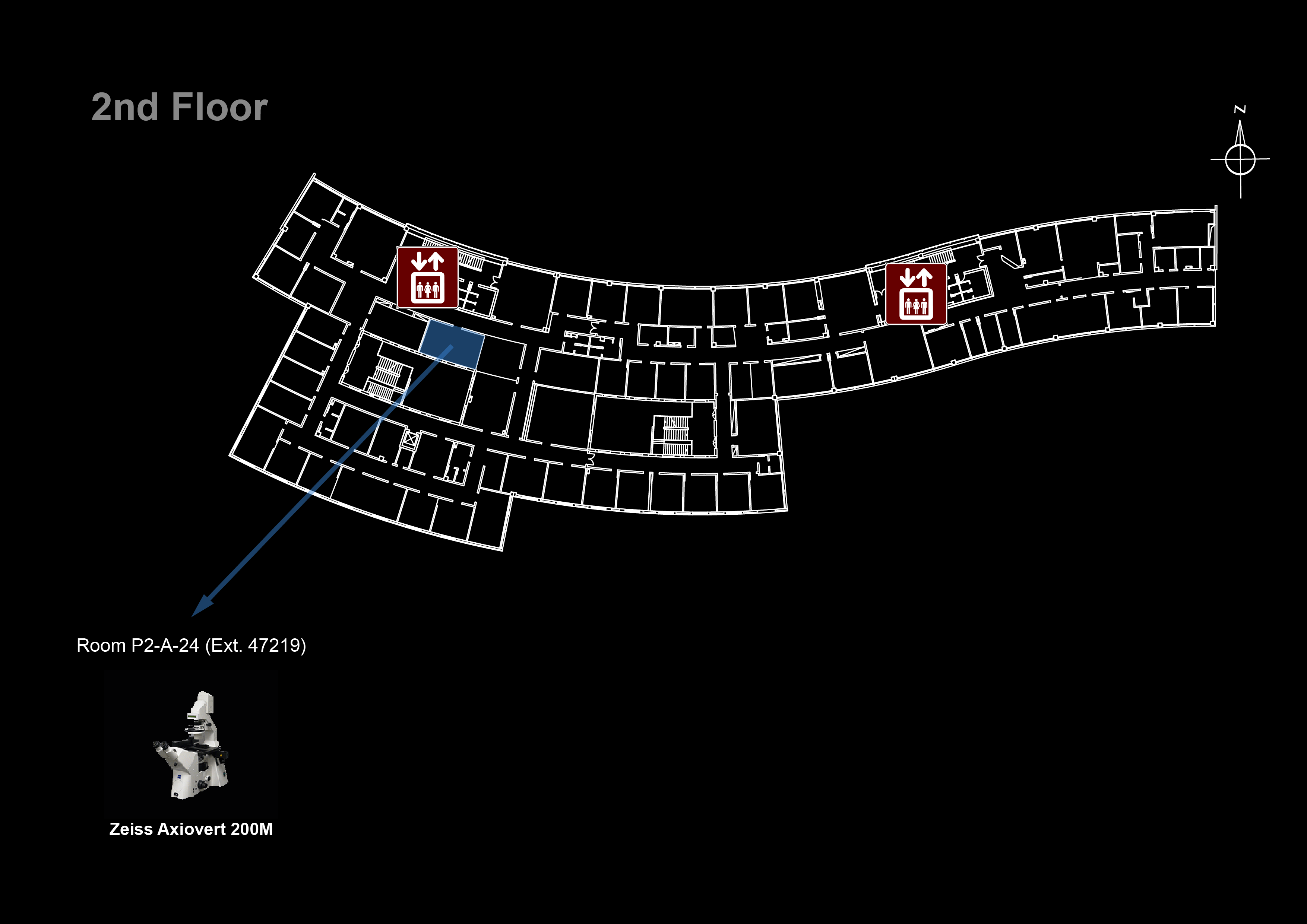
Microscope overview
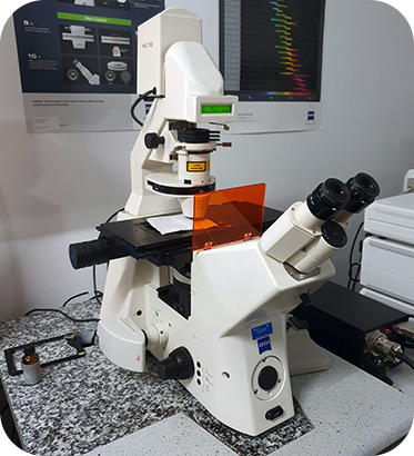
 The Zeiss Axiovert 200M is a fully motorized inverted widefield fluorescence microscope ideal for live-cell imaging applications. Using the Metamorph software you can control the motorized filters, shutters and stage to simultaneously set up multi-color time-lapse imaging experiments in multiple stage positions. Its sensitive cooled CCD camera (Photometrics CoolSNAP HQ CCD) allows you to capture weak fluorescent signals and minimize photobleaching/photodamage in light sensitive samples. You can also perform Z-stack acquisition and use PPBI Huygens RM deconvolution server to significantly improve image resolution even in thick samples. If you are performing long time-lapse experiments in live samples (aqueous media) and need hardware focus control, check the Zeiss Cell Observer with Definite Focus or the Nikon Eclipse Ti. If don't want to use deconvolution but need optical sectioning, check out one of the confocal systems such as the point scanners Zeiss LSM 710 and Zeiss LSM 880 or the spinning disks 3i Marianas SDC and Zeiss Cell Observer SD. If your personal computer cannot handle all the data you collected, check out the Big Guy or Colossus. If you have FISH or other fixed fluorescence slides try the Leica DM5000B instead.
- Microscope: Zeiss Axiovert 200
- Monochrome Camera: Photometrics Coolsnap HQ CCD
- Color Camera: Leica DFC450
![]() Data files older than 1 month will be automatically deleted on this system, please copy your data to the iMM server using the desktop link.
Data files older than 1 month will be automatically deleted on this system, please copy your data to the iMM server using the desktop link.
System components
Filtersets
| Position | Filterset | Reference | Excitation | Dichroic | Emission |
|---|---|---|---|---|---|
| 1 | Blue | FS01 | 359-371 nm | 395 nm | > 397 nm |
| 2 | Green BP | FS10 | 450-490 nm | 510 nm | 515-565 nm |
| 3 | Red | FS20 | 540-552 nm | 560 nm | 575-640 nm |
| 4 | Far Red | Cy5-4040C-000 | 608-648 nm | 660 nm | 672-712 nm |
| 5 | A | none | |||
Upon request:
| Position | Filterset | Reference | Excitation | Dichroic | Emission |
|---|---|---|---|---|---|
| 2 | Green LP | FS09 | 450-490 nm | 510 nm | > 515 nm |
Objectives
| Magnification | Model | Immersion | NA | WD (mm) | pixel size* (μm) | 10 μm = | (color) 10 μm = | Reference |
|---|---|---|---|---|---|---|---|---|
| 10x | EC Plan-Neofluar | Air | 0.30 | 5.20 | 1.00 | 10 pixels | 23.3 pixels | 420340-9900-000 |
| 20x | EC Plan-Neofluar | Air | 0.50 | 2.00 | 0.48 | 21 pixels | 46.7 pixels | 440340-9904-000 |
| 40x | EC Plan-NeoFluar | Air | 0.75 | 0.71 | 0.24 | 41 pixels | 93.7 pixels | 440350-9903-000 |
| 63x | Plan-Apochromat DIC | Oil | 1.40 | 0.19 | 0.15 | 65 pixels | - | 420782-9900-000 |
| 100x | α Plan-Apochromat DIC | Oil | 1.46 | 0.11 | 0.09 | 105 pixels | - | 420792-9800-000 |
Upon request:
| Magnification | Model | Immersion | NA | WD (mm) | pixel size* (μm) | 10 μm = | Reference |
|---|---|---|---|---|---|---|---|
| 1.25x | EC Epiplan-Neofluar | Air | 0.035 | 4 | 7.58 | 1.3 pixels | 422310-9900-000 |
| 5x | EC Plan-NeoFluar | Air | 0.16 | 18.5 | 2.00 | 5 pixels | 420330-9901-000 |
| 40x | Plan-Neofluar Ph3 | Oil | 1.30 | 0.20 | 0.24 | 41 pixels | 440451-9903-000 |
| 63x | Plan-Apochromat Ph3 | Oil | 1.4 | 0.19 | 0.15 | 65 pixels | 420781-9910-000 |
* pixel sizes for binning = 1
Cameras
| Model | Type | Frame Size | Pixel Size (µm) |
|---|---|---|---|
| Photometrics Coolsnap HQ | CCD | 1392 x 1040 | 6.45 x 6.45 |
| Leica DFC450 | color CCD | 2560 × 1920 | 3.4 x 3.4 |
Stage
For automated acquisition of 96 well plates use: X = -9014 μm Y = -8995 μm
System Turn On Procedures
- Turn on the fluorescent light power source

- Turn on the Prior stage controller

- Turn on the microscope

- Turn on the CCD camera

- Turn on the computer
- Log in to Windows (Bioimaging User)
- Start the Metamorph software
- If the software doesn't detect the camera, power cycle the camera and try again
Microscope Turn Off procedures
If there is another user for this microscope in the next hour:
- Leave the fluorescent lamp on
- Clean up immersion objectives
Else:
- Clean up immersion objectives
- Shutdown the computer
- Turn off the camera
- Turn off the Prior stage controller
- Turn off the microscope
- Turn off the fluorescent lamp