User Tools
Table of Contents
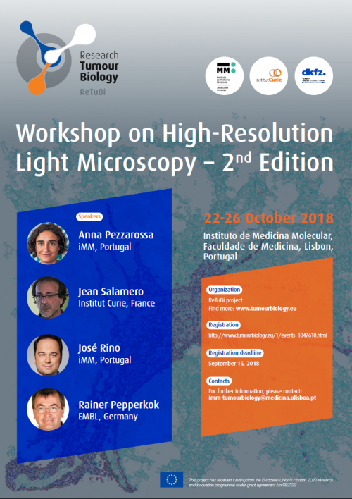
ReTuBi Workshop 2nd edition - 2018
Description
Quantitative fluorescence microscopy has become one of the most important technical advances for the study of cancer progression. The aim of this workshop is to cover the most recent advances in fluorescence microscopy, which enable the observation of cellular activity with unprecedented resolution. The workshop aims at potentiating the participants’ skills in microscopy, help them choosing the most suitable microscope for their particular research question, and learning how to draw quantitative information from their images.
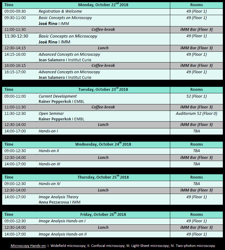
The workshop is organized over 5 days, with 4 lectures of three hours each and 5 hands-on sessions. The participants will have the opportunity to experience directly with four different setups. Each participants will be given a series of lab notes for both the microscopy and the image analysis hands-on to support their
work.
Program
The hands-on sessions will enable the participants to directly experiment with different microscopy techniques. Four different microscopes will be used: widefield, confocal, light-sheet and two-photon microscope. The participants will be divided in four groups of four people to allow everyone enough time to use the microscopes.
There will be two more hands-on session for image analysis, where the participants will have the chance to analyze a set of different images provided by the instructors to answer different questions relative to cell biology and cancer. Mainly, automated cell tracking, and the analysis of FRAP and FRET experiments. Each participant will have the chance to analyze his/her own images and get suggestions and guidance as well as help with the automation of image analysis.
Speakers
- Anna Pezzarossa – iMM, Portugal
- Jean Salamero – Institut Curie, France
- José Rino – iMM, Portugal
- Reiner Pepperkok – EMBL, Germany
Faculty for labs
- Ana Nascimento – iMM
- Anna Pezzarossa – iMM
- António Temudo – iMM
- José Marques - iMM
- José Rino – iMM
Equipment
- Zeiss LSM 880 | point-scanning confocal microscope
- Leica SP8 MP | multiphoton fluorescence microscope
- Zeiss Lightsheet Z1 | lightsheet fluorescence microscope
- Zeiss Cell Observer | widefield fluorescence microscope
Registration
Registrations are already closed. Thank you for your interest.
Further Information
For further course information please contact José Rino at joserino@medicina.ulisboa.pt


