
This is an old revision of the document!
Table of Contents

|
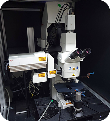
Location: Room P2-B-42 ( |
 Leica SP8 MP Booking
 Leica SP8 MP Usage Statistics
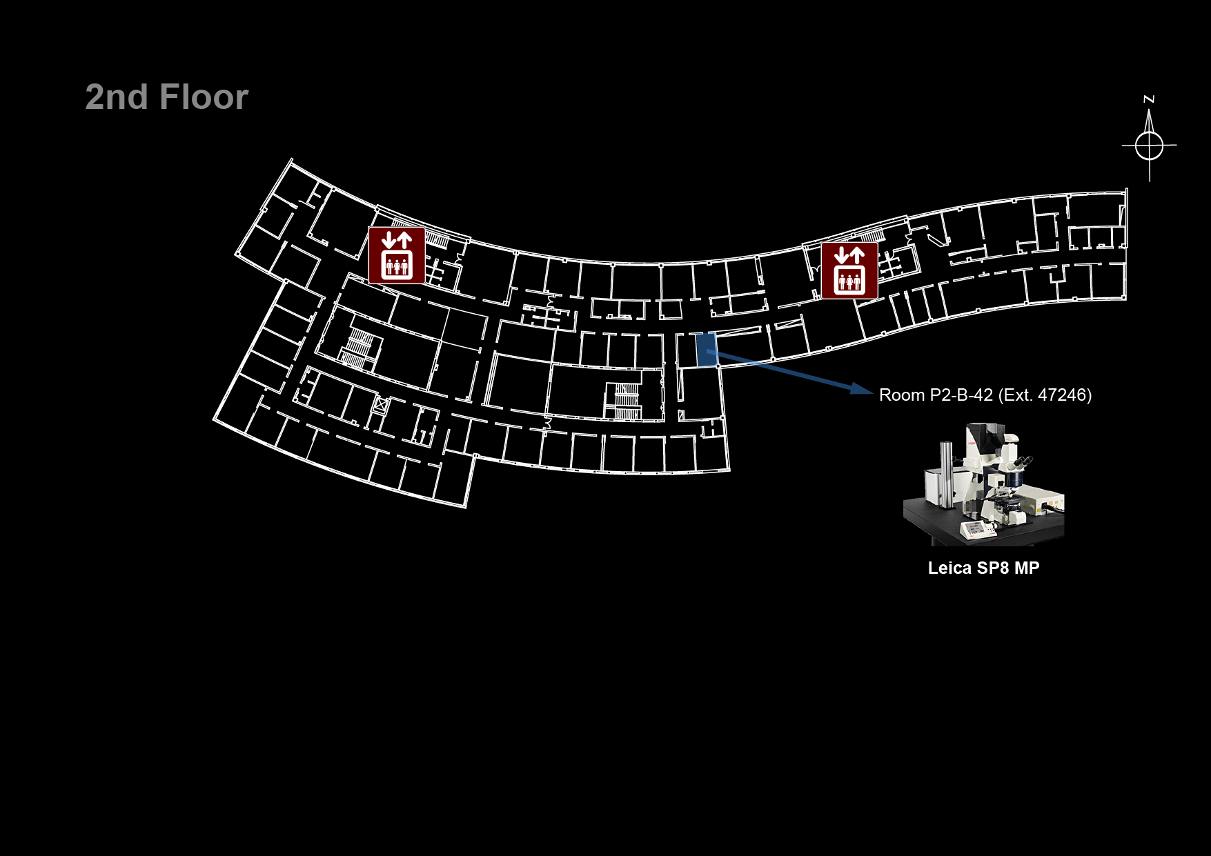
Microscope overview
 The Leica SP8 MP is a both a confocal and multi-photon microscope able to generate high-resolution three-dimensional images of thick specimens. Multi-photon excitation (most commonly two photon excitation) is particularly advantageous for imaging thicker samples. Rather than exciting the fluorophore with one photon, multi-photon excitation is produced by two or more lower energy photons which can penetrate deeper in the sample. Moreover, contrarily to confocal, the multi-photon excitation light will only achieve sufficient intensity to cause fluorescence in a specific region. Because of this, no pinhole is needed in multi-photon microscopy to exclude the light from out-of-focus planes and achieve optical sectioning. The Leica SP8 MP is an upright microscope equipped with water and glycerol immersion objectives especially suitable for intra-vital and thick samples immersed in water or glycerol imaging. In confocal mode, its scanning unit includes a spectral detector PMT to be used with a 488 nm laser for excitation. In multi-photon mode, its Insight DS+ Dual pulsed laser can be tuned from 680 to 1300 nm and has a second laser line emitting at 1041 nm, to be used with four non-descanned detectors with specific filtersets: two PMTS and two HyD detectors (hybrid detectors with 45% QE compared to ~25% QE for conventional PMT). With this system you can perform optical sectioning high resolution imaging of fluorescent samples that are two thick for confocal microscopes such as the Zeiss LSM 880 or the Zeiss LSM 710, albeit with a slightly lower resolution. If you do not need optical sectioning and your sample is thin, check out a widefield system instead, such as the Cell Observer or the Nikon Eclipse Ti. If your personal computer cannot handle all the data you collected, check out the Big Guy or Colossus.
![]() Click on the image on the right to see it in higher detail
Click on the image on the right to see it in higher detail
![]() Data files older than 3 months will be automatically deleted on this system, please copy your data to the iMM server using the desktop link.
Data files older than 3 months will be automatically deleted on this system, please copy your data to the iMM server using the desktop link.
System components
LASERs
| Laser Unit | Wavelength | Maximum Power | Current Status |
|---|---|---|---|
| InSight DS+ Dual (multi-photon) | 680 - 1300 nm | 1.3 W | not working |
| 1041 nm | 1.5 W | not working | |
| SS OBIS 488-20 (confocal) | 488 nm | 20 mW | ok |
Objectives
| Magnification | Model | Immersion | NA | WD (mm) | Reference |
|---|---|---|---|---|---|
| 25x | HC FLUOTAR VISIR | Water | 0.95 | 2.5 | 15506374 |
| 63x | HC PL APO Glyc CORR CS2 | Glycerol | 1.30 | 0.30 | 15506353 |
Filtersets (Ocular)
| Filterset | Reference | Excitation | Emission | Fluorochromes |
|---|---|---|---|---|
| Blue | I3 | 450-490 nm | > 515 nm | GFP, FITC, Alexa488 |
Emission Filters (multi-photon)

| Detector | Filter | Transmission | Fluorochromes |
|---|---|---|---|
| HyD-RLD 1 | Red | 570-640 nm | mCherry, Alexa568, Cy3 |
| HyD-RLD 2 | Green | 500-550 nm | GFP, Alexa488, FITC |
| PMT-RLD 3 | Far Red | 662 - 737 nm | Alexa647, Cy5 |
| PMR-RLD 4 | Red | 570-640 nm | mCherry, Alexa568, Cy3 |
Upon request:
(requires changing filters and the dichroic beamsplitter)

| Detector | Filter | Transmission | Fluorochromes |
|---|---|---|---|
| HyD-RLD 1 | Green | 500-550 nm | GFP, Alexa488, FITC |
| HyD-RLD 2 | Blue | 415-485 nm | DAPI, Hoescht, Alexa350 |
| PMT-RLD 3 | Far Red | 662 - 737 nm | Alexa647, Cy5 |
| PMR-RLD 4 | Red | 570-640 nm | mCherry, Alexa568, Cy3 |
System Turn On Procedures
- Check that the cooling and power supply for the infrared laser are ON (
InSight DeepSee READY)

- Turn on the fluorescence lamp

- Turn on the HyD (Hybrid detectors) supply unit

- Turn on the computer
- Turn on the microscope electronics box

- Turn on the microscope's control panel

- Switch on the scanner power, laser power and activate the key switch to turn on the 488 nm laser

- Log in to Windows (Bioimaging User)
- Start the LAS X software