
This is an old revision of the document!
Table of Contents

|
Location: Room P2-A-22 ( |
 Zeiss LSM 880 Booking
 Zeiss LSM 880 Usage Statistics
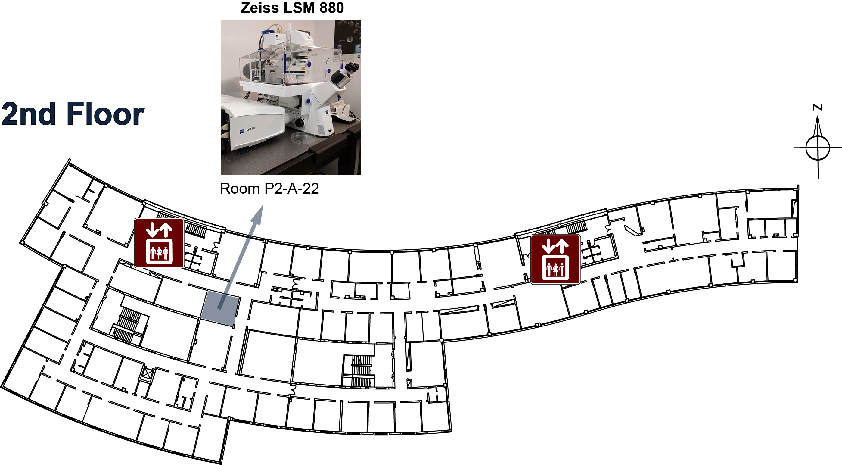
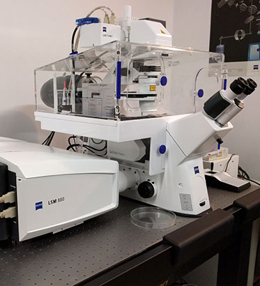
Microscope overview
The Zeiss LSM 880 with Airyscan is a confocal point-scanning microscope able to generate high-resolution three-dimensional images of thick specimens with high sensitivity and low photodamage. It is an inverted microscope specially suitable for live cell imaging and photobleaching experiments, equipped with a large size incubator for temperature control and CO2 supply. Its Airyscan 32 channel area detector is able to collect all light from an Airy pattern simultaneously, allowing for increased resolution (140 nm laterally and 400 nm axially, at 488 nm). Its scanning unit further includes two PMT detectors (one of them cooled for single photon counting) as well as a GaAsP detector. It is equipped with lasers from violet to far red (405, 458, 488, 561 and 633 nm excitation wavelengths). With this system you can perform optical sectioning of fluorescent samples which are too thick for a widefield system such as the Zeiss Axiovert 200M. Image resolution and detection sensitivity are higher than faster confocal systems such as a spinning disk or the Zeiss LSM 7 Live, but image acquisition is slower. If you need to use a 594 nm laser or a 40x water objective for higher working distances, check the Zeiss LSM 510 META confocal point-scanning system. If your personal computer cannot handle all the data you collected, check out the Big Guy.
System components
LASERs
| Laser Unit | Wavelength | Maximum Power | Current Status |
|---|---|---|---|
| Sapphire 488-100 | 488 nm | 100 mW | ok |
| DPSS 561-40 | 561 nm | 40 mW | ok |
Objectives
| Magnification | Model | Type | NA | WD (mm) |
|---|---|---|---|---|
| 10x | W Achroplan Ph1 | Water dipping | 0.30 | 3.1 |
| 10x | Plan-Neofluar | Dry | 0.30 | 5.5 |
| 20x | W Plan-Apochromat | Water | 1.00 | 1.8 |
| 40x | W Achroplan | Water dipping | 0.80 | 3.6 |
| 40x | Plan-Neofluar Ph3 | Oil | 1.30 | 0.20 |
Filtersets (Ocular)
| Position | Filterset | Reference | Excitation | Emission |
|---|---|---|---|---|
| 2 | Green | FS10 | 450-490 nm | 515-565 nm |
| 3 | Red | FS15 | 540-552 nm | > 590 nm |
| 4 | Blue (for ablation*) | FS49 | none | 420-470 nm |
* Excitation filter has been removed
Filter and Dichroic sets
 |  |
| Main configuration | Emission filters |
Microscope Turn On Procedures
Normal Operation
- The system must be turned on 30 min before usage.
- To start it turn on the two main switches:
SYSTEM/PCandCOMPONENTS

- Login to the computer, wait for the network icon to change to
connectedstate - Start the ZEN 2009 software.
After the 30 min:
- Turn on the metal halide lamp (white box to the left of the microscope).

- You may now start using the system.
Live Imaging / Time Lapse Operation
- The system must be turned on 2 hours before usage (in order to properly warm up scanning mirrors).
- To start it turn on the two main switches: SYSTEM/PC and COMPONENTS
- Login to the computer, wait for the network icon to change to
connectedstate - Start the ZEN 2009 software.
After the 2 hours:
- Turn on the metal halide lamp (white box to the left of the microscope).
- You may now start using the system.
Microscope Turn Off procedures
If there's another user for this microscope in the next two hours:
- Exit the ZEN 2009 software, leave the lasers on and log off the computer.
- Clean up immersion objectives.
- Make sure that there really is another user going to use the microscope.
Else:
- Turn off the lasers, wait 5 min for them to cooldown.
- Exit the ZEN 2009 software and shut down the computer.
- Clean up immersion objectives.
- Turn off the metal halide lamp.
- Turn off the microscope.
- Turn off the two main switches: SYSTEM/PC and COMPONENTS
If you want to use the UV laser ablation system:
- Please contact the Bioimaging Facility (Ext: 47316). The ablation system requires specific training even for users that are already trained on the LSM 7 Live itself.