
This is an old revision of the document!
Table of Contents

|
Location: Room P2-A-24 ( |
 Zeiss Celldiscoverer 7 Usage Statistics
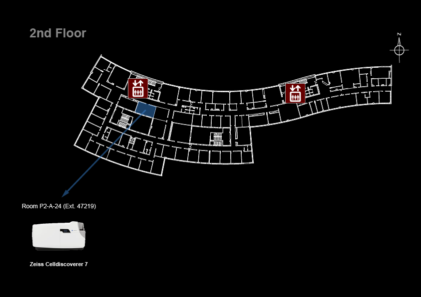
page under construction
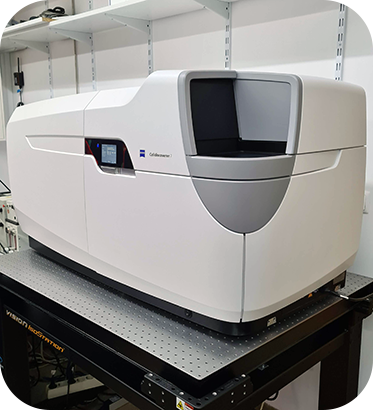
Microscope overview
 The Zeiss Celldiscoverer 7 is a fully integrated and motorized high-end inverted widefield fluorescence imaging system ideal for automated live-cell imaging in multi-well plates and Petri dishes. It offers a fully integrated incubation system with temperature and CO2 control and Definite Focus.3 for hardware focus control. The ZEN software can automatically calibrate the system and adapt if to your sample by recognizing different sample carriers, determining their bottom thickness and material and adjusting the objectives correction ring to compensate for spherical aberrations. You can use Petri dishes, chamber slides, multi-well plates, plastic or glass, thin or thick vessel bottoms, low-skirt or high-skirt plates. When using the 50x water immersion objective, automated immersion supply and removal is available for time lapse experiments over several days or extensive scanning processes on multi-well plates. The fluorescence excitation unit combines seven LEDs – from deep blue to far-red - for high-speed multi-channel imaging. Its sensitive cooled Axiocam 702 and 712 cameras allows you to capture weak fluorescent signals and minimize photobleaching/photodamage in light sensitive samples. If you need optical sectioning for thick samples, check out one of the confocal systems such as the point scanners Zeiss LSM 880 or Zeiss LSM 980 or spinning disks 3i Marianas SDC and Zeiss Cell Observer SD. If your personal computer cannot handle all the data you collected, check out the Colossus.
- LED Illumination: Zeiss Colibri 7
- Cameras: Axiocam 702 mono CMOS and Axiocam 712 mono CMOS
![]() Data files older than 1 month will be automatically deleted on this system, please copy your data to the iMM server using the desktop link.
Data files older than 1 month will be automatically deleted on this system, please copy your data to the iMM server using the desktop link.
System components
Filtersets
| Position | Filterset | Reference | Dichroic | Emission |
|---|---|---|---|---|
| 1 | RQBS | FS90HE | 405 nm | 410-440 nm |
| 493 nm | 499-529 nm | |||
| 575 nm | 580-605 nm | |||
| 653 nm | 659-759 nm | |||
| 2 | RTBS | FS91HE | 450 nm | 455-479 nm |
| 538 nm | 543-568 nm | |||
| 610 nm | 615-760 nm | |||
| 3 | RTBS | FS92HE | 405 nm | 410-440 nm |
| 493 nm | 499-549 nm | |||
| 610 nm | 616-761 nm | |||
| 4 | RDBS | FS93HE | 493 nm | 498-530 nm |
| 575 nm | 580-630 nm |
Objectives
| Magnification | Model | Immersion | NA | WD (mm) | Afocal magnification |
|---|---|---|---|---|---|
| 2.5x | |||||
| 5x | EC Plan-Neofluar Ph1 | Air | 0.30 | 5.20 | 2.5x |
| 20x | Plan-Apochromat | Air | 0.80 | 0.55 | 420650-9901-000 |
| 20x | EC Plan-Neofluar Ph 2 | Air | 0.50 | 2.00 | 440341-9904-000 |
| 40x | EC Plan-NeoFluar | Air | 0.75 | 0.71 | 420360-9900-000 |
| 63x | Plan-Apochromat DIC | Oil | 1.40 | 0.19 | 420782-9900-000 |
| 63x | Plan-Apochromat Ph3 | Oil | 1.40 | 0.19 | 420781-9910-000 |
Camera
| Model | Type | Frame Size | Pixel Size (µm) |
|---|---|---|---|
| Axiocam 506 mono | CCD | 2752 x 2208 | 4.54 x 4.54 |
System Turn On Procedures
- Turn on the HXP 120 fluorescent light source (if used)

- Turn on the temperature and CO2 controller - white power strip (if you need them).
- If you need CO2, open the Cell Observer CO2 valve
- Turn on the motorized stage controller (A on remote control)

- Turn on the microscope power supply (B on remote control)

- Turn on the computer
- Login in to Windows with your Agendo credentials
- Start the ZEN software
- Select HXP or LED Workspace in ZEN (top left)

Microscope Turn Off procedures
If there is another user for this microscope is the next hour:
- Leave the fluorescent light source on
- Clean up immersion objectives
Else:
- Exit the ZEN software
- Turn off the computer (not the UPS!)

- Clean up immersion objectives
- Turn off the microscope power supply (B on remote control)
- Turn off the motorized stage controller (A on remote control)
- Turn off the Temperature and CO2 controller - white power strip (if used)
- Close the Cell Observer CO2 valve (if used)
- Turn off the HXP 120 fluorescent light source (if used)