
Table of Contents

|
Location: Lisbon Site, Room P2-A-49 ( |
 Cytek Amnis ImageStreamX Booking
 Cytek Amnis ImageStreamX Usage Statistics
 Cytek Amnis ImageStream Brochure
System overview
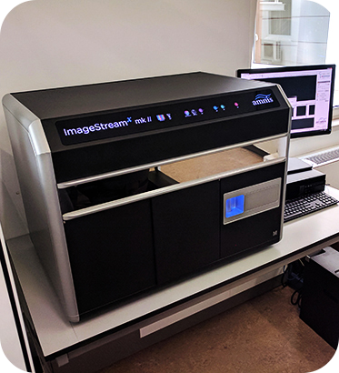
 The Cytek Amnis ImageStreamX is a benchtop multispectral imaging flow cytometer equipped with two CCD cameras that can acquire up to 12 images simultaneously of each cell or object in brightfield, scatter and multiple fluorescent channels at rates up to 5000 objects/sec. It has five lasers - 375 nm, 488 nm, 561 nm, 642 nm and 785 nm (dedicated to side scatter). Cells in suspension are hydrodynamically focused in a flow cell, where they are illuminated by a brightfield light source and by lasers. High numerical aperture (NA) objective lenses collect fluorescence emission, as well as scattered and transmitted light from the cells which then intersect a spectral decomposition element that focus different spectral bands on different physical locations of two CCD cameras. By collecting large numbers of digital images per sample and providing numerical representation of image-based features, the ImageStreamX combines single cell standard microscopy information with the statistical significance of large sample sizes in standard flow cytometry. While you can use it to measure fluorescence intensity as in conventional flow cytometers, the best applications for the ImageStreamX take advantage of its imaging abilities to locate and quantify the distribution of signals within cells or even between cells in cell conjugates. The system also has a dedicated software application used for spectral compensation, image analysis and statistical analysis of acquired images (IDEAS) with predefined wizards for apoptosis, cell cycle, co-localization, internalization, shape change, spot counting and others.
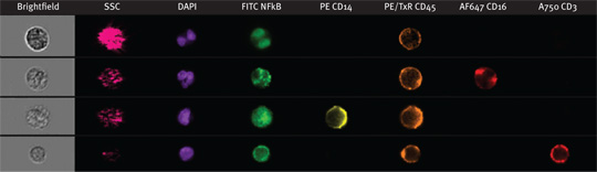
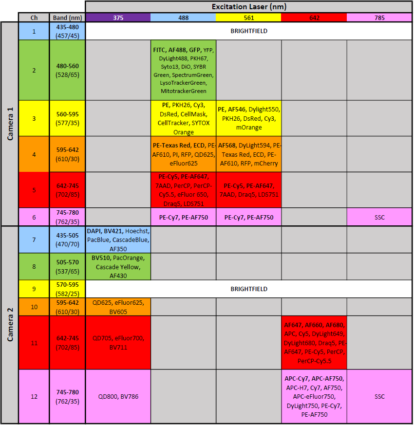
- 12 imaging channels (2 brightfield channels + 10 channels for fluorescence detection)
- 5 lasers with UV (375 nm), blue (488 nm), yellow-green (561 nm), red (642 nm) and far red (785 nm) for side scatter
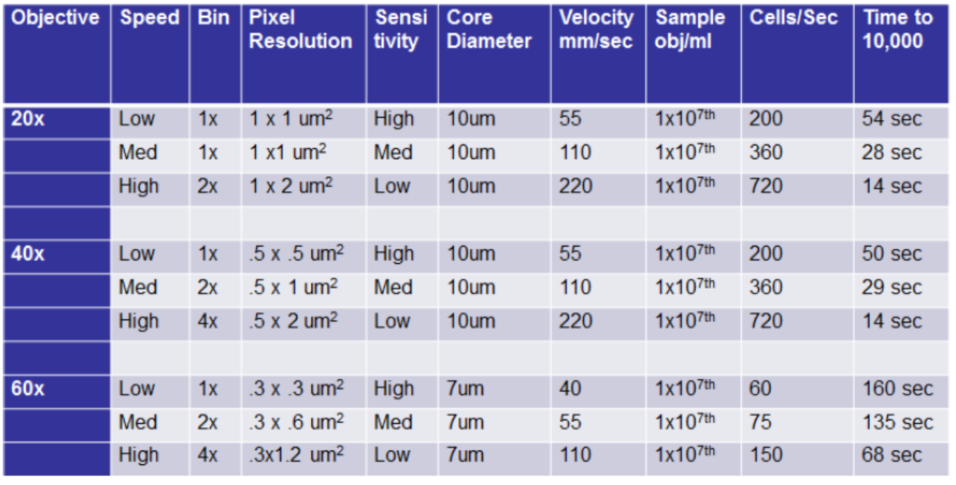
- Multimag: 20x, 40x and 60x magnification
- Extended depth of field: projects all structures within a cell into a single plane of focus
Configuration
Objectives Information
| Magnification | 20x | 40x | 60x |
|---|---|---|---|
| Numerical Aperture | 0.50 | 0.75 | 0.90 |
| Field of View (μm) | 120 | 60 | 40 |
| Pixel Size (μm) | 1.0 | 0.5 | 0.33 |
| Depth of Field (μm) | 8.0 | 4.0 | 2.5 |
Image Acquisition Performance
Additional information

- Sample Preparation Guide. Note: Samples are run using 1.5 ml microfuge tubes (Sarstedt ref. 72706)
Booking Rules
Available 7 days a week, 24 hours a day for trained researchers.
Please check the User Guidelines for detailed information.