User Tools
Table of Contents

|
Location: [Oeiras] Room 0B02 |
 ZEISS Axio Imager with Apotome 2 Usage Statistics
![[Oeiras] Room 0B02](/facility/bioimaging/lib/exe/fetch.php?media=bartolomeu_dias_wing_2025_-_apotome.png)
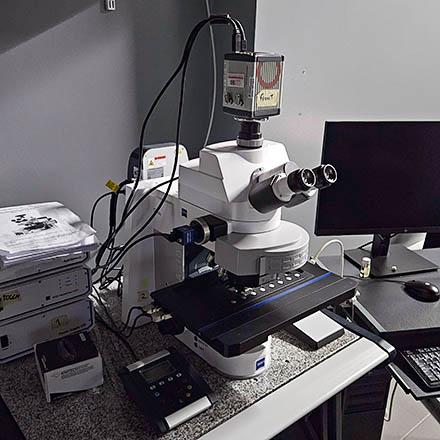
Microscope overview
The ZEISS Axio Imager with Apotome 2 is a fully automated upright microscope capable of magnifications from 2.5x to 160x and equipped with two cameras: On the left port a 5MPx CMOS color camera (Axiocam 105 RGB, for brightfield/DIC) and on the top port a high sensitivity 4 Mpx monochrome sCMOS camera (Hamamatsu Flash 4.0v2, for fluorescence). The system has a Colibri 7 excitation light source with 5 LEDs (see table below). The system’s stage, which can accommodate 8 slides simultaneously, is motorized in XYZ, capable of z-stacks, multiple positions or tile and stitching acquisition (thereby allowing acquisition of large fields of view with several hundred megapixels). The Imager is also equipped with ApoTome.2, which allows optical sectioning of fluorescent samples using structured illumination. A grid pattern is placed on the illumination path and is projected and shifted sideways; 3 (or more) images are acquired and between each image the grid is moved 1/3 of its cycle. This movement generates images where only the in-focus light is modulated by the grid pattern while out-of-focus light is largely unaffected by the grid. By subtracting pairs of these images, the system removes out-of-focus light and the grid pattern, thus creating an optical section of the sample.
System components
LEDs
| Line | Wavelength | Power (with 10x) |
|---|---|---|
| UV | 385 nm | ~ 160* mW |
| B | 475 nm | ~ 120 mW |
| G | 555 nm | ~ 20 mW |
| Y | 590 nm | ~ 14 mW |
| R | 630 nm | ~ 44 mW |
* don't exceed 5% power as this line bleaches samples quickly.
Filtersets
| Filterset | Excitation | Dichroic | Emission | Reference |
|---|---|---|---|---|
| Blue | 370-410 nm | 420 nm | 430-470 nm | FS96HE |
| Green | 450-490 nm | 493 nm | 500-550 nm | FS38HE |
| Orange-Red | 540-552 nm | 580 nm | > 590 nm | FS15 |
| Red | 625-655 nm | 660 nm | 665-715 nm | FS50 |
| DIC | - | - | - | - |
| Orange | 575-600 nm | 605 nm | 612-682 nm | FS64HE |
| QUAD | 370-400 nm | 405 nm | 410-440 nm | FS90HE |
Objectives
| Magnification | Model | Immersion | NA | WD (mm) | Reference |
|---|---|---|---|---|---|
| 2.5x | Plan-Neofluar | air | 0.085 | 8.8 | 420320-9902-000 |
| 10x | Plan-Neofluar | air | 0.3 | 5.5 | 420340-9900-000 |
| 20x | Plan-Apochromat | air | 0.8 | 0.55 | 420650-9902-000 |
| 40x | Plan-Apochromat | air | 0.95 | 0.25 | 420660-9970-000 |
| 40x | Plan-Neofluar | oil | 1.3 | 0.21 | 420462-9900-000 |
| 100x | Plan-Apochromat | oil | 1.4 | 0.17 | 420792-9900-000 |
Camera
| Model | Frame Size | Pixel Size (µm) | Quantum Efficiency |
|---|---|---|---|
| Hamamatsu ORCA-Flash4.0 V2 | 2048 x 2048 | 13.3 x 13.3 | 70 % |
| ZEISS Axiocam 105 color | 2048 x 2048 | 2.2 x 2.2 | - |
Microscope Turn On Procedure
- Turn on the Power Supply

- Turn on the Microscope Body

- Turn on the computer
- Log In into Windows with your Agendo credentials
- Start the ZEN software
Microscope Turn Off Procedure
If there is another user for this microscope in the next hour:
- Exit the ZEN software
- Log off the computer
- Clean up immersion objectives
Else:
- Exit the ZEN software
- Clean up immersion objectives
- Turn off the Microscope Body
- Turn off the Power Supply
- Turn off the computer