This is an old revision of the document!
Table of Contents

|
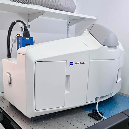
Location: Room 0B03 ( |
 UPDATE Zeiss Lightsheet Z.1 Usage Statistics
Microscope overview
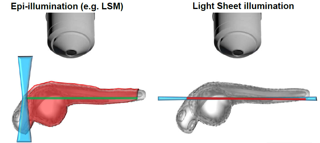
 The Zeiss Lightsheet Z.1 is a light sheet fluorescence microscope able to image optical sections of large samples at subcellular resolution and very fast rates, with almost no phototoxicity or bleaching. It splits fluorescence excitation and detection into two separate light paths, with the axis of illumination being perpendicular to the detection axis.
Since only a single thin section of the sample is illuminated by the light sheet, optical sectioning is achieved without a pinhole or image processing deconvolution. Light from the in-focus plane is collected by a sCMOS camera rather than pixel by pixel as in a point scanning laser confocal. This allows you to collect images faster and with less excitation light than you would with many other optical-sectioning microscopy techniques. This system features two objectives with fields of view around 1 mm or 500 µm and resolutions between 0.15 µm and 0.3 µm, equipped with lasers spanning blue to far-red wavelengths (445 nm, 488 nm, 561 nm, and 638 nm) and appropriate fluorescence emission filters. Designed for live imaging, samples are embedded in agarose and preferably loaded into glass or plastic capillaries that provide heating and cooling to maintain physiological conditions, making the system ideal for observing live cells, embryos, Drosophila, and zebrafish. It is important to note that this Z.1 is optimized for live samples and does not include a kit for cleared samples; for such applications, other microscopes are recommended. Users should consult with facility staff to determine the best technique for their specific research needs.
The Zeiss Lightsheet Z.1 is a light sheet fluorescence microscope able to image optical sections of large samples at subcellular resolution and very fast rates, with almost no phototoxicity or bleaching. It splits fluorescence excitation and detection into two separate light paths, with the axis of illumination being perpendicular to the detection axis.
Since only a single thin section of the sample is illuminated by the light sheet, optical sectioning is achieved without a pinhole or image processing deconvolution. Light from the in-focus plane is collected by a sCMOS camera rather than pixel by pixel as in a point scanning laser confocal. This allows you to collect images faster and with less excitation light than you would with many other optical-sectioning microscopy techniques. This system features two objectives with fields of view around 1 mm or 500 µm and resolutions between 0.15 µm and 0.3 µm, equipped with lasers spanning blue to far-red wavelengths (445 nm, 488 nm, 561 nm, and 638 nm) and appropriate fluorescence emission filters. Designed for live imaging, samples are embedded in agarose and preferably loaded into glass or plastic capillaries that provide heating and cooling to maintain physiological conditions, making the system ideal for observing live cells, embryos, Drosophila, and zebrafish. It is important to note that this Z.1 is optimized for live samples and does not include a kit for cleared samples; for such applications, other microscopes are recommended. Users should consult with facility staff to determine the best technique for their specific research needs.
![]() Click on the image on the right to see the system beam path in higher detail
Click on the image on the right to see the system beam path in higher detail
![]() Data files older than 1 month will be automatically deleted on this system, please copy your data to the file server using the desktop link.
Data files older than 1 month will be automatically deleted on this system, please copy your data to the file server using the desktop link.
Additional information for sample preparation
- Sample Size: up to 1 x 1 x 2 cm
- Penetration depth: up to 5.6 mm
Tutorials for Image Analysis with Arivis
System components
LASERs
| Laser Unit | Wavelength | Maximum Power |
|---|---|---|
| Solid State 488 | 488 nm | 30 mW |
| Solid State 561 | 561 nm | 20 mW |
| Solid State 638 | 638 nm | 75 mW |
Objectives (Illumination)
| Magnification | Model | NA |
|---|---|---|
| 10x | Lightsheet Z.1 10x | 0.20 |
Objectives (Detection)
| Magnification | Model | Immersion | NA | WD (mm) | Reference |
|---|---|---|---|---|---|
| 20x | W Plan-Apochromat DIC 75mm | Water | 1.00 | 2.4 | 421452-9700-000 |
| 20x | Clr Plan-Neofluar Corr nd=1.45 85mm | Clearing | 1.00 | 5.6 | 421459-9970-000 |