
Table of Contents

|
Location: Room P2-B-42 ( |
 Leica SP8 MP Usage Statistics
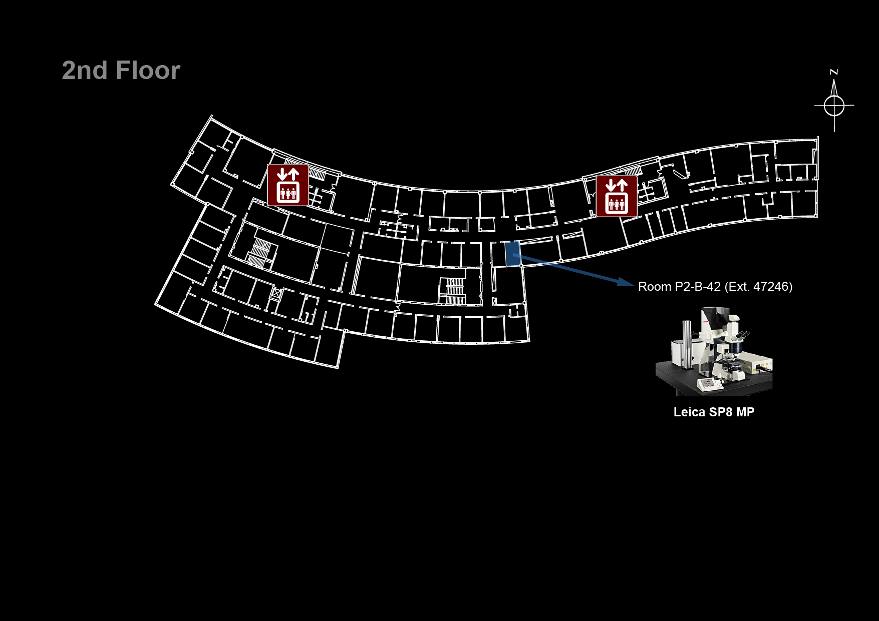
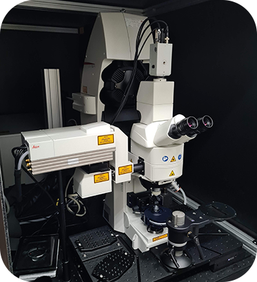
Microscope overview
 The Leica SP8 MP is a both a confocal and multi-photon microscope able to generate high-resolution three-dimensional images of thick specimens. Multi-photon excitation (most commonly two photon excitation) is particularly advantageous for imaging thicker samples. Rather than exciting the fluorophore with one photon, multi-photon excitation is produced by two or more lower energy photons which can penetrate deeper in the sample. Moreover, contrarily to confocal, the multi-photon excitation light will only achieve sufficient intensity to cause fluorescence in a specific region. Because of this, no pinhole is needed in multi-photon microscopy to exclude the light from out-of-focus planes and achieve optical sectioning. The Leica SP8 MP is an upright microscope equipped with water and glycerol immersion objectives especially suitable for intra-vital and thick samples immersed in water or glycerol imaging. In confocal mode, its scanning unit includes a spectral detector PMT to be used with a 488 nm laser for excitation. In multi-photon mode, its Insight DS+ Dual pulsed laser can be tuned from 680 to 1300 nm and has a second laser line emitting at 1041 nm, to be used with four non-descanned detectors with specific filtersets: two PMTS and two HyD detectors (hybrid detectors with 45% QE compared to ~25% QE for conventional PMT).
![]() Click on the image on the right to see it in higher detail
Click on the image on the right to see it in higher detail
![]() Data files older than 3 months will be automatically deleted on this system, please copy your data to the iMM server using the desktop link.
Data files older than 3 months will be automatically deleted on this system, please copy your data to the iMM server using the desktop link.
System components
LASERs
| Laser Unit | Wavelength |
|---|---|
| InSight DS+ Dual (multi-photon) | 680 - 1300 nm |
| 1040 nm | |
| SS OBIS 488-20 (confocal) | 488 nm |
Objectives
Filtersets (Ocular)
| Filterset | Reference | Excitation | Dichroic | Emission | Fluorochromes |
|---|---|---|---|---|---|
| Blue | I3 | 450-490 nm | 510 nm | > 515 nm | GFP, FITC, Alexa488 |
Emission Filters (multi-photon)

| Detector | Filter | Transmission | Fluorochromes |
|---|---|---|---|
| HyD-RLD 1 | Red | 570-640 nm | mCherry, Alexa568, Cy3 |
| HyD-RLD 2 | Green | 500-550 nm | GFP, Alexa488, FITC |
| PMT-RLD 3 | Far Red | 662 - 737 nm | Alexa647, Cy5 |
| PMR-RLD 4 | Red | 570-640 nm | mCherry, Alexa568, Cy3 |
Upon request:
(requires changing filters and the dichroic beamsplitter)

| Detector | Filter | Transmission | Fluorochromes |
|---|---|---|---|
| HyD-RLD 1 | Green | 500-550 nm | GFP, Alexa488, FITC |
| HyD-RLD 2 | Blue | 415-485 nm | DAPI, Hoescht, Alexa350 |
| PMT-RLD 3 | Far Red | 662 - 737 nm | Alexa647, Cy5 |
| PMR-RLD 4 | Red | 570-640 nm | mCherry, Alexa568, Cy3 |
System Turn On Procedures
- Check that the cooling and power supply for the infrared laser are ON (
InSight DeepSee READY)

- Turn on the fluorescence lamp

- Turn on the HyD (Hybrid detectors) supply unit

- Turn on the computer
- Turn on the microscope electronics box

- Turn on the microscope control panel

- Switch on the scanner power, laser power and turn the key switch to on

- Login in to Windows with your Agendo credentials
- Start the LAS X software
Microscope Turn Off procedures
If there is another user for this microscope in the next hour:
- Leave the fluorescent lamp and lasers on
- Clean up immersion objectives
Else:
- Clean up immersion objectives
- Set the infrared laser to
hibernate(deactivate the infrared laser in the Currently Available Lasers window) - Close the LAS X software
- Turn of the 488 nm laser with the key switch in the flexible supply unit
- Switch off the HyD supply unit
- Switch off the microscope control panel
- Shutdown the computer
- Switch off the laser power and scanner power in the flexible supply unit
- Turn off the microscope electronics box
- Turn off the fluorescent lamp