This is an old revision of the document!
Table of Contents

|
Location: [Oeiras] Room 0B05 |
 Andor Dragonfly Usage Statistics
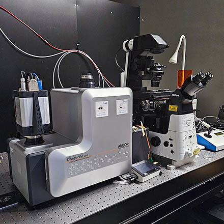
Microscope overview
Spinning disk confocal microscopy (SDC) is a technique well suited for observation of cells and organoids. Compared to conventional widefield microscopy, SDC provides instant optical sectioning, allowing the imaging of cells and shallow tissues (typically a maximum of a few tens of micrometres deep) in 3D. Compared to laser scanning confocal microscopes (such as Leica Stellaris, Zeiss Airyscan), the Dragonfly SDC provides much faster acquisition (up to 400 fps, with limited ROI (2048×128), 40 fps full frame), with lower phototoxicity and photobleaching. A spinning disk with multiple small pinholes is installed between the light source and specimen to generate point-like illumination covering the whole area “simultaneously”. Each small aperture serves as a detecting pinhole to remove out-of-focus fluorescence. This system was built to be versatile, and allow imaging of different sample sizes. It includes both high and low magnification objectives (which require different pinhole sizes, 25 or 40um). The scanhead allows 1x or 2x magnification (for extra magnification and to allow Nyquist sampling and deconvolution - see table below).
System components
| Position | Filterset | Reference | Excitation | Dichroic | Emission |
|---|---|---|---|---|---|
Objectives
| Magnification | Model | Immersion | NA | WD (mm) | Reference |
|---|---|---|---|---|---|