User Tools
This is an old revision of the document!
Table of Contents


|
Location: [Oeiras] Room 0B02 |
Microscope overview
GABY WORKING ON IT!!!!
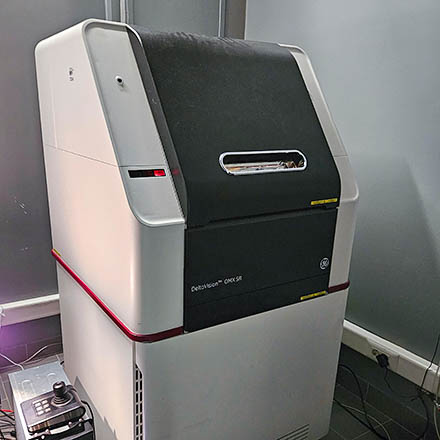
SIM microscopy is one of the imaging techniques that can overcome the limits of resolution in a normal widefield microscope (~200nm), by using patterned (“structured”) illumination with sliding and rotating stripes. These patterns produce high order spatial information that can later be extracted by computational algorithms in Fourier space. A typical SIM image is a reconstruction of several patterned images, where the illumination pattern changes in both angle or phase in order to extract almost twice the original resolution. in the DV OMX system, a total of 15images are acquired for each image (5x phases x3 angles). SIM works best with shallow samples, such as attached cells, and is less employed for tissue imaging. Optimal optical conditions are important to avoid reconstructions artifacts. When successful, SIM can provide valuable information about organelles, cytoskeleton and fine intra cellular details. The speed of acquisition of the DV OMX system also allows live-cell imaging.
Suggestion for description in “Materials and Methods”: “Images were acquired on an Deltavision OMX system, equipped with 2 PCO Edge 5.5 sCMOS cameras, using the a 60x 1.42NA Oil immersion objetive, GFP + CY5 fluorescence filtersets and DIC optics. Images were deconvolved/reconstructed with Applied Precision's softWorx software.
System components
Lasers
| Excitation | Wavelength | Power |
|---|---|---|
| Violet | 405 nm | 100mW |
| Blue | 488 nm | 100mW |
| Green | 568 nm | 100mW |
| Red | 640 nm | 100mW |
Objectives
| Magnification | Immersion | NA | WD (mm) | Image Pixel Size | Reference |
|---|---|---|---|---|---|
| 20x UPlan SAPO | - | 0.75 | 0.6 | 0.240 | |
| 60x Plan APO N | Oil | 1.42 | 0.15 | 0.080 |
"Channels" & respective emission filters/cameras
| Position | Emission wavelength | Dyes | Camera |
|---|---|---|---|
| DAPI | 435/31 | DAPI, CFP, Hoescht… | 1 |
| GFP | 528/48 | GFP, Alx488… | 1 |
| RFP | 609/37 | RFP, Alx568… | 2 |
| Cy5 | 683/40 | GCy5, Alx633… | 2 |
| DIC | - | Brighfield | 1/2 |
Cameras (1 and 2)
2x PCO Edge 5.5 sCMOS 2560×2160 (note that each camera uses only 1024×1024 pixels to avoid field illumination problems and to allow simultaneous acquisition of 2 channels per camera)
 Deltavision OMX-SIM Usage Statistics