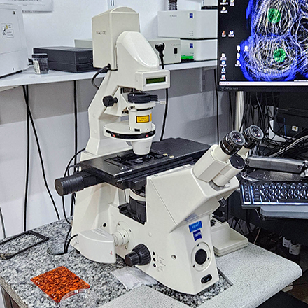
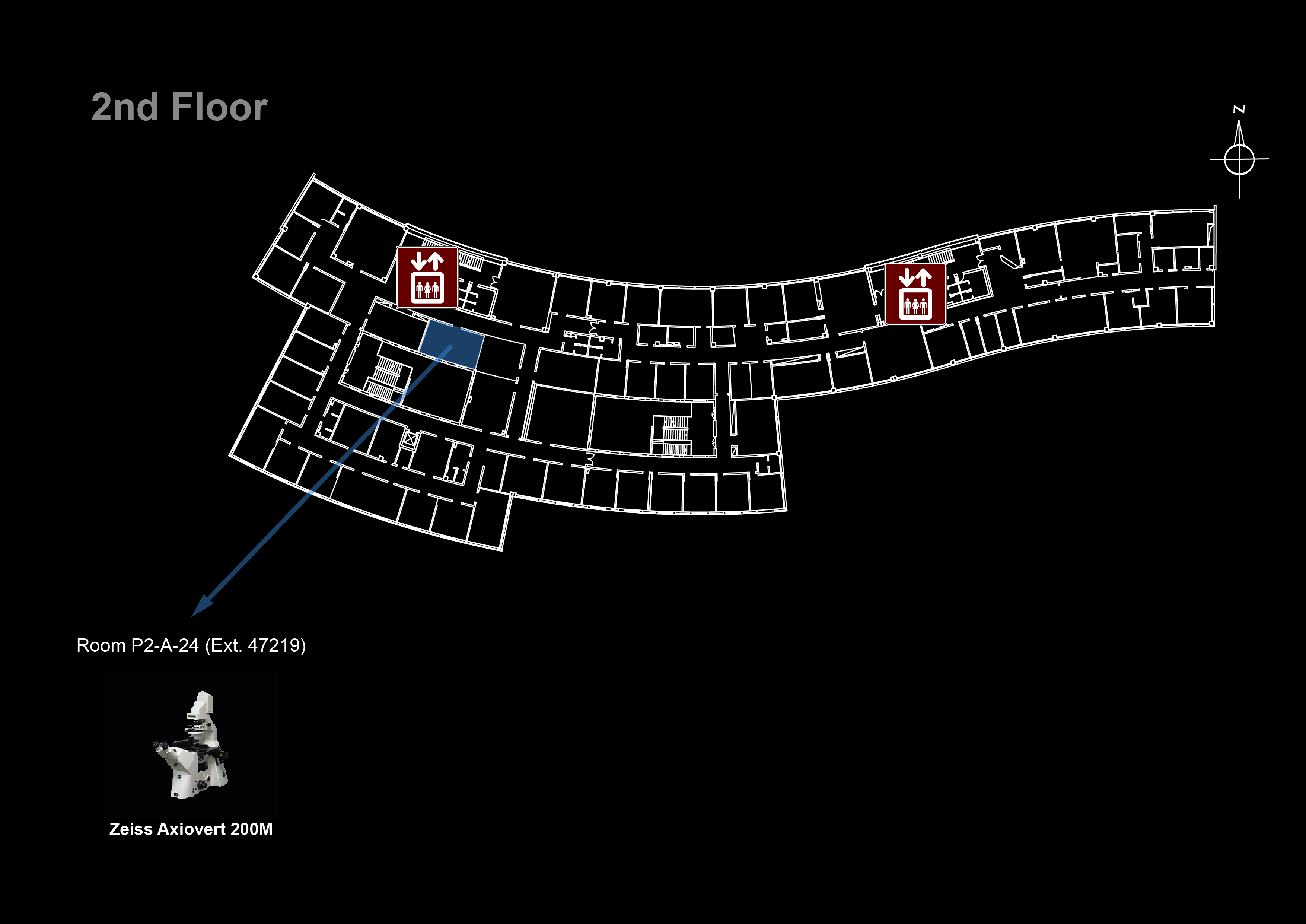
Location: [Lisbon] Room P2-A-24 ( 47219)
Manufacturer: ZEISS Microscopy
Model: Axiovert 200M
Nickname: "Axiovert"
Software: Metamorph 7.8.0.0
Year: 2003
SN: ZCA522000494
 
Data will be deleted after: 1 month  
 
→ Quality Control
→ Usage Statistics
===== Microscope overview ===== The Zeiss Axiovert 200M is a fully motorized inverted //widefield fluorescence microscope// ideal for live-cell imaging applications. Using the Metamorph software you can control the motorized filters, shutters and stage to simultaneously set up multi-color time-lapse imaging experiments in multiple stage positions. Its sensitive cooled CCD camera ([[https://labs.pbrc.edu/cellbiology/documents/CoolSNAPHQ.pdf|Photometrics CoolSNAP HQ CCD]]) allows you to capture weak fluorescent signals and minimize photobleaching/photodamage in light sensitive samples. It is also equipped with a color camera ([[https://www.leica-microsystems.com/products/microscope-cameras/p/leica-dfc450-c/|Leica DFC450]]) for acquisition of histological stains with tile and stitching imaging. * **Microscope**: Zeiss Axiovert 200 * **Monochrome Camera**: [[https://labs.pbrc.edu/cellbiology/documents/CoolSNAPHQ.pdf|Photometrics Coolsnap HQ CCD]] * **Color Camera**: [[https://www.leica-microsystems.com/products/microscope-cameras/p/leica-dfc450-c/|Leica DFC450]] {{:warning.png?25|}} Data files older than **1 month** will be automatically deleted on this system, please copy your data to the GIMM server using the desktop link. ===== System components ===== ==== Filtersets ====
Position Filterset Excitation Dichroic Emission Reference
1 Blue 359-371 nm 395 nm > 397 nm FS01
2 Green BP 450-490 nm 510 nm 515-565 nm FS10
3 Red 540-552 nm 560 nm 575-640 nm FS20
4 Far Red 608-648 nm 660 nm 672-712 nm Cy5-4040C-000
5 none
**Upon request:**
Position Filterset Excitation Dichroic Emission Reference
2 Green LP 450-490 nm 510 nm > 515 nm FS09
==== Objectives ====
Magnification Model Immersion NA WD (mm) pixel size* (μm) 10 μm = (color) 10 μm = Reference
10x EC Plan-Neofluar Air 0.30 5.20 1.00 10 pixels 23.3 pixels 420340-9900-000
20x EC Plan-Neofluar Air 0.50 2.00 0.48 21 pixels 46.7 pixels 440340-9904-000
40x EC Plan-NeoFluar Air 0.75 0.71 0.24 41 pixels 93.7 pixels 440350-9903-000
63x Plan-Apochromat DIC Oil 1.40 0.19 0.15 65 pixels - 420782-9900-000
100x α Plan-Apochromat DIC Oil 1.46 0.11 0.09 105 pixels - 420792-9800-000
**Upon request:**
Magnification Model Immersion NA WD (mm) pixel size* (μm) 10 μm = Reference
1.25x EC Epiplan-Neofluar Air 0.035 4 7.58 1.3 pixels 422310-9900-000
5x EC Plan-NeoFluar Air 0.16 18.5 2.00 5 pixels 420330-9901-000
40x Plan-Neofluar Ph3 Oil 1.30 0.20 0.24 41 pixels 440451-9903-000
63x Plan-Apochromat Ph3 Oil 1.4 0.19 0.15 65 pixels 420781-9910-000
* **pixel sizes for binning = 1** ==== Cameras ====
Model Type Frame Size Pixel Size (µm)
Photometrics Coolsnap HQ CCD 1392 x 1040 6.45 x 6.45
Leica DFC450 color CCD 2560 × 1920 3.4 x 3.4
==== Stage ==== For automated acquisition of 96 well plates use: X = -9014 μm Y = -8995 μm ===== Turn ON Procedure ===== * Turn on the fluorescent light power source (if used) {{halide_710.jpg?80|}} * Turn on the Prior stage controller {{ax_prior_on.jpg?100|}} * Turn on the microscope {{ax_mic_on.jpg?100|}} * Turn on the CCD camera {{ax_cam_on.jpg?100|}} * Turn on the computer (if needed) * Log in to Windows (Bioimaging User) * Start the Metamorph software * If the software doesn't detect the camera, power cycle the camera and try again ===== Turn OFF procedure ===== If there is another user for this microscope in the __next hour__: * Leave the fluorescent lamp on (if used) * Clean up immersion objectives Else: * Clean up immersion objectives * Turn off the computer * Turn off the camera * Turn off the Prior stage controller * Turn off the microscope * Turn off the fluorescent lamp (if used) ---- [[resources|Back to the Equipment page]]